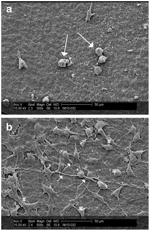
Figure 7. Effect of laminin on neuron growth.
DRG neurons were plated on aerogel surfaces and grown for 7 days (to confluence) prior to processing for SEM. (a) SEM of nerve growth profile off the laminin dot, directly on the aerogel substrate. Arrows indicating attached nerve cells with no processes extended. (b) SEM of DRGs grown on a laminin drop on PCSA disc post-fixation and sputtering. DRGs cell bodies (*) are extending processes ( ) that interact with those of other nerves. Sample processing (freezing and sputter-coating) has lead to the fracturing of the lamimin layer (#). Images taken post-fixation and sputtering stage.

