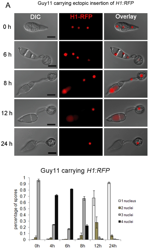
Figure 1. Nuclear degeneration occurs during appressorium development in M. oryzae.
(A). Upper panel: time course live cell imaging experiment showing nuclear division and nuclear degeneration during appressorium development in M. oryzae. Guy11 conidia expressing H1:RFP were examined by epifluorescence microscopy at indicated time points during appressorium development. Lower panel: bar charts showing the percentage of spore germlings in Guy11 containing between 0 and 4 nuclei (mean ± SD, n>100, triple replications) during a timecourse of appressorium development. Scale bar = 10 µm.

