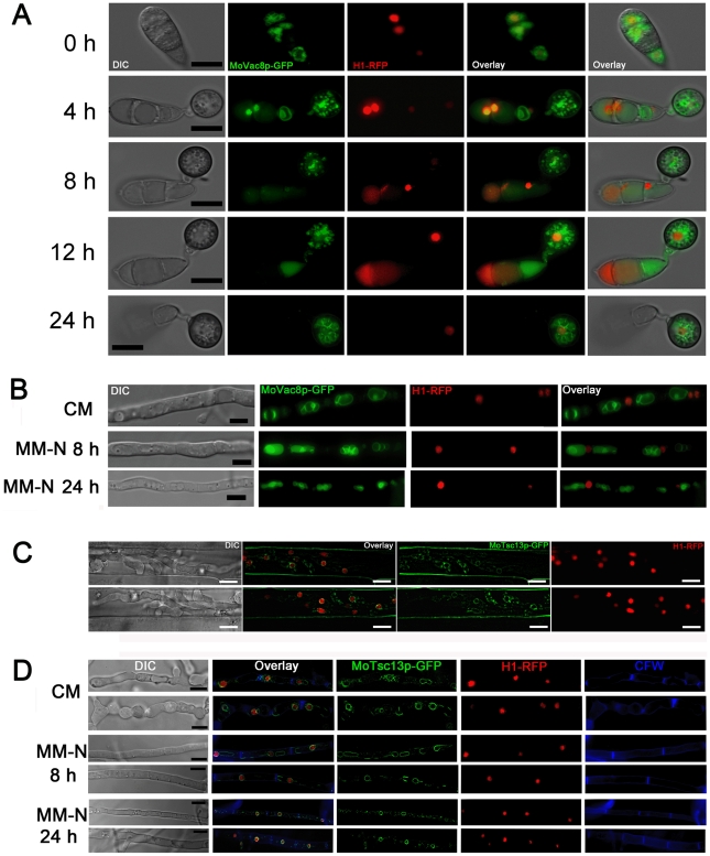
Figure 6. Nuclear-vacuolar (NV) junctions are not present in M. oryzae during appressorium development or starvation stress.
(A) Time course live cell imaging of appressorium development in M. oryzae expressing both MoVAC:GFP and H1:RFP. (B) Micrographs of hyphae of M. oryzae expressing both MoVAC:GFP and H1:RFP. M. oryzae was grown in nutrient-rich medium (CM) or nitrogen stress minimal medium (MM-N) for the indicated period of times, and hyphae collected for epifluorescence microscopy. Hyphae cultured in CM for 40 h were washed with distilled water, before being transferred into MM-N. (C) MoTsc13p is associated with perinuclear and peripheral ER membrane in invasive hyphae. M. oryzae expressing both MoTSC13:GFP and H1:RFP was inoculated onto rice sheath epidermis for 36 h before epifluorescence microscopy. (D) Distribution of MoTsc13p-GFP is not affected by environmental nutritional status. Hyphae of M. oryzae expressing both MoTSC13:GFP and H1:RFP were grown in CM or MM-N for epifluorescence microscopy. Pictures shown are representatives of at least 30 different hyphae cell compartments analyzed for each time point. Scale bar = 10 µm.

