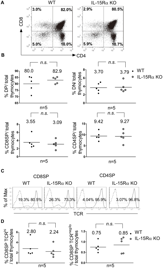
Figure 1. Characterization of thymocyte subpopulations in WT and IL-15Rα KO mice.
Total thymocytes were isolated from WT and IL-15Rα KO mice (n = 5 per group) and subjected to flow cytometric analysis of the expression of CD4, CD8, and TCRβ. (A) Thymic subsets of WT and IL-15Rα KO mice were shown for their expression of CD4 and CD8. (B) Percentages of DN, DP, CD4SP, and CD8SP thymocytes among total thymocytes of IL-15Rα KO (○) mice and wild-type littermates (•) were compared. The horizontal line represents the mean. (C) CD4SP and CD8SP thymocytes were further gated to compare their TCR expression. CD8SP thymocytes consisted of TCRneg/lo and TCRhi cells, whereas the majority of CD4SP cells expressed high levels of TCR. (D) The percentage of CD8SP TCRhi cells (left panel) and CD8SP TCRneg/lo cells (right panel) among total thymocytes was compared between WT and IL-15Rα KO mice. Data were analyzed by single-classification ANOVA. n.s., not significant.

