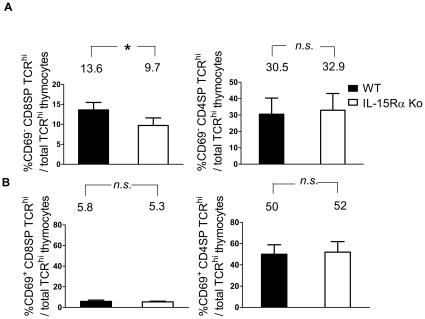
Figure 2. Examination of CD69-negative SP thymocytes in WT and IL-15Rα KO mice.
Thymocytes of WT and IL-15Rα KO mice (n = 6) were immunostained for CD4, CD8, TCR, and CD69. The percentages of CD69-negative (A) and CD69-positive (B) CD8SP TCRhi (left panels) and CD4SP TCRhi thymocytes (right panels) among total TCRhi cells were compared. By calculation, the cell numbers of CD69− CD8SP TCRhi thymocytes in WT and IL-15Rα KO mice were about 1.6×10⁶±3.8×10⁵ and 9.0×10⁵±2.8×10⁵, respectively. The cell numbers of CD69− CD4SP TCRhi thymocytes in WT and IL-15Rα KO mice were about 3.6×10⁶±1.3×10⁵ and 3.2×10⁶±1.4×10⁵, respectively. Data are presented as means ± SD and were analyzed by single-classification ANOVA. *p<0.05; n.s., not significant.

