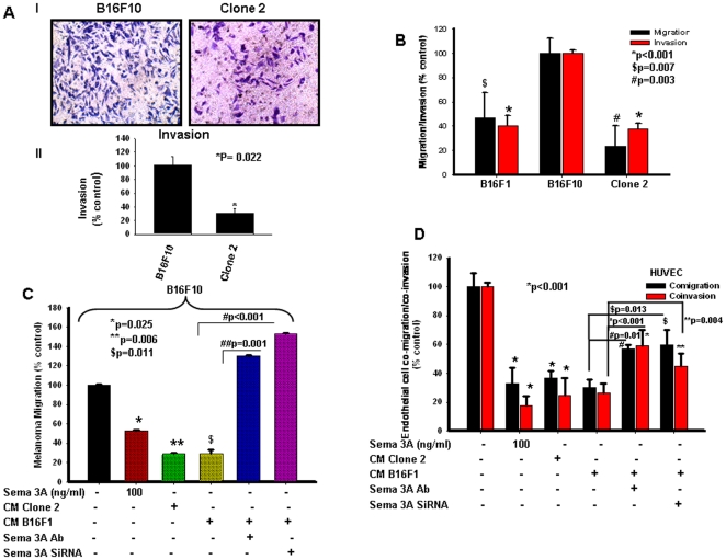
Figure 3. Sema 3A inhibits melanoma cell migration and invasion through autocrine and paracrine mechanisms.
(A) Invasion assays were performed on matrigel coated invasion chamber. Cells (control or clone 2; 1×10⁵) were plated on upper chamber. After 18 h, invaded cells were stained with Giemsa and photographed (panel I), counted in 3 high-power fields (C/HPF) under an inverted microscope (Nikon), analyzed statistically and represented in the form of bar graph (panel II, *P = 0.022). (B) Autocrine mechanism of Sema 3A action on melanoma motility and invasiveness were demonstrated by migration and invasion assay using either B16F10 or clone 2 or B16F1 cells. *p<0.001, $p = 0.007, #p = 0.003 vs. B16F10. (C) Boyden chamber migration assay was performed using B16F10 cells to demonstrate the paracrine mechanism of action of Sema 3A. Conditioned media (CM) collected from clone 2 or B16F1 cells alone or B16F1 transfected with Sema 3A siRNA were used in the lower chamber as a chemoattractant. In separate experiments, either B16F10 cells treated with Sema 3A (100 ng/ml) or CM from B16F1 treated with Sema 3A antibody were used in the lower chamber. Migrated cells were counted, analyzed statistically and represented graphically. *p = 0.025, **p = 0.006 and $p = 0.011 vs. control, #p<0.001, ##p = 0.001. (D) Tumor-endothelial interaction was performed in modified Boyden chamber or matrigel coated invasion chamber. Endothelial cells were placed on upper chamber whereas similar conditioned media (as of Fig. 3C) were used in the lower chamber. Migrated or invaded cells were stained with Giemsa, photographed, analyzed statistically and represented in the form of graph. *p<0.001 vs. control, **p = 0.004, #p = 0.01, $p = 0.013.

