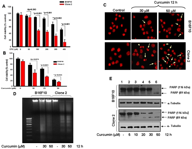
Figure 5. Sema 3A overexpressed clone exhibits increased drug sensitivity in B16F10 cells.
(A) Control or clone 2 cells were treated with melanoma specific drug, Dacarbazine (DTIC) (0–400 µM) for 24 h and cells survival was analyzed by MTT assay. #p = 0.393, *p<0.001, **p = 0.002 vs. respective treatment group within B16F10 and clone 2 cells. (B) Similarly, both cells were treated with curcumin (0–50 µM) for 12 h and cell viability was checked by MTT assay. The data are represented in the form of bar graph and the mean value of triplicate experiments is indicated. #p = 0.074, *p<0.001, **p = 0.044 vs. respective treatment group within B16F10 and clone 2 cells. (C) Both cells were treated with indicated concentrations of curcumin, fixed and stained with PI (red) and photographed under fluorescence microscope at 60× magnifications. The apoptotic nuclei are indicated by arrows. (D) Cells were treated with two doses of curcumin for 12 h. Fragmentation of genomic DNA was extracted and resolved on 2% agarose gel. Apoptotic DNA fragmentation was visualized by ethidium bromide staining. (E) Curcumin induced apoptosis in control and clone 2 cells were also analyzed by Western blot using anti-PARP antibody. Cells were treated with 0–50 µM curcumin for 12 h and then analyzed by Western blot. α-Tubulin was used as loading control. The experiments showed here are representative of triplicate independent experiments with similar results.

