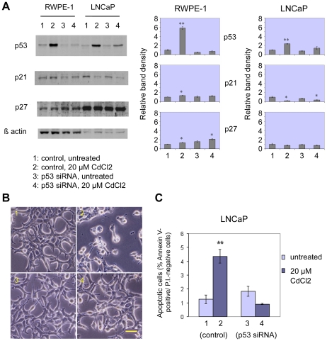
Figure 4. Silencing of p53 suppresses apoptosis induction by cadmium in human prostate cells.
p53 siRNA transfection: western blot analysis (RWPE-1 and LNCaP cells) and early apoptosis detection (LNCaP cells). WB analysis of p53, p21 and p27 expression (A), representative phase contrast microscopy images (B) and FITC-conjugated Annexin-V/PI and FACS analysis (C; histograms, reporting mean percentages ± SEM of Annexin-V positive/PI-negative cells, n = 3) after p53 siRNA transfection, followed by 24-h treatment with 20 µM CdCl2. In LNCaP cells siRNA-mediated p53 silencing is able to suppress apoptosis induction by 24-hour exposure to 20 µM cadmium chloride, as compared to the respective control. WB histograms represent relative band densities (mean ± SEM, n = 3), as determined by densitometry, using β-actin as the loading control for standardization. Scale bar in B: 100 µm. *P<0.05, **P<0.01.

