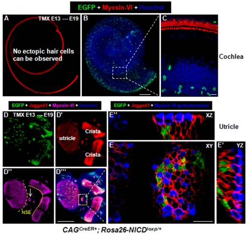
Figure 5. Overactivation of NICD at E13 generates new HCs in the utricle but not the cochlea.
(A–C) Whole-mount cochlear image of a CAGCreER+; Rosa26-NICDloxp/+ embryo treated with tamoxifen at ∼E13 and analyzed at ∼E19. Although many EGFP+ cells were present, no new HCs were observed. (D–E″) Whole-mount images of the utricle and 2 adjacent cristae from the same embryo. White arrows in (D″) point to the ectopic sensory epithelia region. (E–E″) A confocal three-dimensional, high-magnification image of the white rectangular region in (D′″). NSE: non-sensory region; XY: Confocal XY plane; YZ: Confocal YZ plane; XZ: Confocal XZ plane. Scale bars: 200 µm in (B and D′″) and 20 µm in (E).

