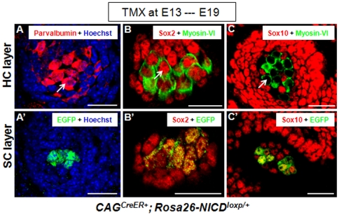
Figure 6. Expression of the sensory epithelium marker Parvalbumin and Sox2 in new HCs, and Sox10 in new SCs.
(A–A′) Images of samples double stained with Parvalbumin and EGFP at HC layer (A) and SC layer (A′) of the ectopic sensory patches in the utricle non-sensory region of a CAGCreER+; Rosa26-NICDloxp/+ embryo treated with tamoxifen at ∼E13 and analyzed at ∼E19. The arrow points to a new Parvalbumin+ HC. (B–B′) Triple staining of Myosin-VI, Sox2, and EGFP. Both ectopic HCs (B) and SCs (B′) were Sox2+. The arrow points to a new Sox2+/Myosin-VI+ HC. Of note, Myosin-VI was visualized in a pseudo-green color. (C–C′) Triple staining of Myosin-VI, Sox10, and EGFP. The arrow points to a new Myosin-VI+/Sox10−negative HC. The SCs (either EGFP+ or EGFP−negative) were Sox10+. Note that Myosin-VI was also visualized in a pseudo-green color. Scale bars: 20 µm.

