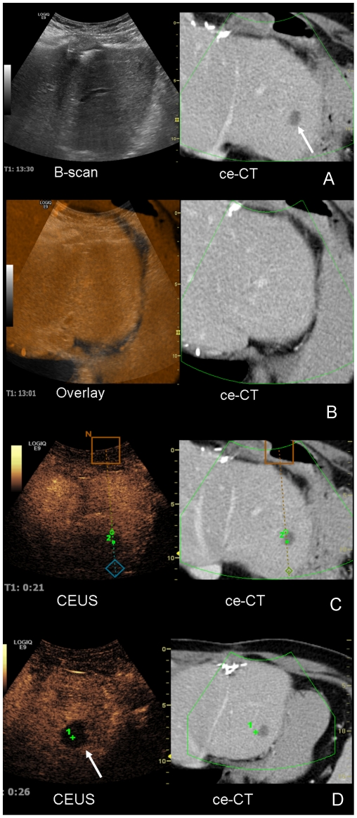
Figure 2. Image fusion (ultrasound and CT) for interventional planning for local radiofrequency ablation.
A 67 years old patient with colorectal carcinoma and several partial liver resections in his history showed a new solitary liver metastasis in segment II of the liver, clearly visible in ceCT. The referring surgeons requested a local radiofrequency ablation of the metastasis. Figure 2 A. The metastasis cannot be detected in fundamental B-scan, but in the ceCT on the right side. Figure 2 B. For image fusion the contrast enhanced CT scan is color-coded and superimposed onto the fundamental B-scan. Figure 2 C. CEUS clearly shows the metastasis, and is therefore used for planning of the radiofrequency ablation. Figure 2 D. CEUS control after radiofrequency ablation with point registration shows complete necrosis in the area of the former metastasis with a safety margin of over 1 cm in all directions.

