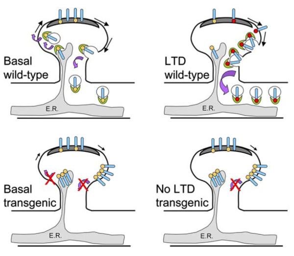
Figure 1.

Schematic of Effects of In Vivo Blockade of PICK1-GluR2 Interaction
Wild-type: induction of LTD results in PICK1 (green crescent) mediated internalization of PKCα phosphorylated Ser880 (red dot) GluR2-containing AMPARs (blue cylinder) from the extrasynaptic spine plasma membrane into the dendrite. There is basal phosphorylation (orange dot) of GluR2 at Ser880 mediated by a kinase other than PKCα. PICK1 does not recruit PKCα to GluR2; rather, the BAR domain of PICK1 is involved in sensing or creating the membrane curvature required for vesicle formation. PICK1 may also be necessary for the export of new AMPARs from the ER (gray shaded region). Representation of forward traffic has been omitted in the LTD diagram for clarity. Transgenic animals: in animals where the PICK1-GluR2 interaction is prevented, there is no change in the number of AMPARs in the PSD (dark gray). However, there is an increase in surface AMPARs on the spine outside the PSD and a decrease in AMPARs in the dendritic shaft presumably because AMPARs fail to internalize from the extrasynaptic spine plasma membrane in the absence of GluR2-PICK1 interactions. The build up of AMPARs inside the spine reflects a role for PICK1 in exit of GluR2-containing AMPARs from the ER. There is no LTD because AMPAR internalization is PICK1 dependent.
