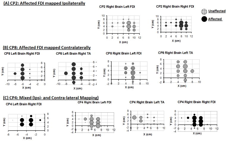
Figure 2.
Examples of cortical motor maps for 3 representative subjects with right hemiplegia. Three patients with the same clinical classification of right hemiplegia showed 3 different corticomotor projection patterns: ipsilateral (A), contralateral (B), and bilateral (C). Note that TA MEPs are evoked at cortical sites >4 cm lateral to the vertex (B and C). Also note the proximity between map locations for FDI and TA (B and C) and for right and left FDI (A and C).

