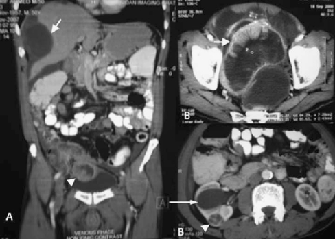
Fig. 1.
Panel A—CECT abdomen showing irregular thick walled peripherally enhancing polypoidal lesion abutting the caecum and terminal ileum with solid-cystic components and infiltration and projection into the urinary bladder (arrow head). Perihepatic high density fluid collections and marked scalloping of the liver (long arrow). Panel B (upper panel)—Pelvic scans showing large cystic peripherally enhancing lesion abutting the rectum (white arrow). Panel B (lower panel)—Dilatation of renal pelvis of the right kidney (long arrow) with a small round well defined hypodense peripherally enhancing lesion with spoke wheel central scar in the lower pole (arrow head)

