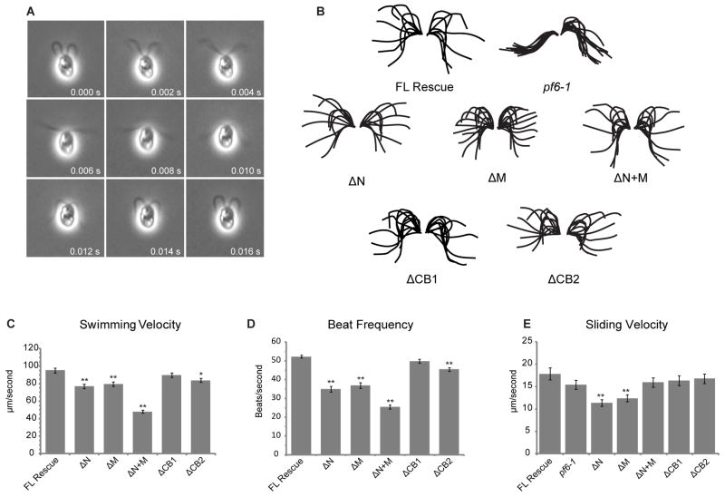
Figure 4. Swimming velocity, beat frequency, and microtubule sliding velocity are reduced in some PF6Δaa strains.
(A) Montage of images obtained from high speed video recording of cell transformed with full-length PF6, demonstrating a complete beat cycle. Videos of all other transformants are included in supplemental data. (B) Tracings of complete beat cycles from pf6-1 and each of the PF6 transformants. pf6 is paralyzed, however, waveforms of all PF6Δaa transformants are similar to flagella containing full-length PF6. Each flagellar trace represents one frame of video separated by 2 ms. (C) Mean swimming velocity of strains containing truncated PF6 protein. Velocity is significantly decreased in PF6ΔN, PF6ΔM, and PF6ΔN+M (**p < 0.001, Student’s t test). A small but significant decrease is also observed in PF6ΔCB2 (*p < 0.01, Student’s t test). (D) Asymmetric beat frequency is significantly decreased in PF6ΔN, PF6ΔM, PF6ΔN+M, and PF6ΔCB2 (**p < 0.001, Student’s t test). (E) Mean microtubule sliding velocities for PF6 strains. Microtubule sliding velocities were obtained using an in vitro microtubule sliding assay and isolated axonemes as indicated in Methods. Velocity is significantly decreased in PF6ΔN and PF6ΔM (**p < 0.001, Student’s t test). N ≥ 50 for all swimming velocity, beat frequency, and sliding velocity measurements. Total N is derived from three independent experiments. Error bars indicate SEM. Data is presented in tabular form in Table 2.

