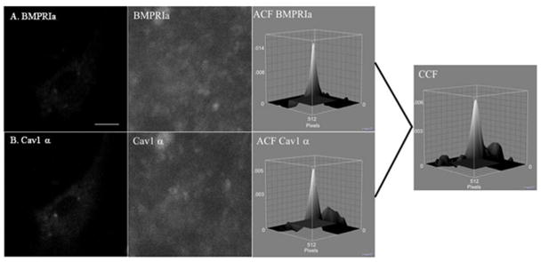
Figure 2. Auto and Cross Correlation Functions of BMPRIa and Cav1 α in C57BL/6J.
BMSCs were isolated and cultured on glass cover slips. The proteins, BMPRIa and Cav1 α, were labeled with a primary followed by a fluorescent secondary antibody. Images were taken at high resolution of the cellular membrane. Following confocal microscopy ICS and ICCS was performed on the images. A sample image of BMSC isolated from the C57BL/6J labeled for A) BMPRIa and B) Cav1 α with the corresponding high resolution image and auto correlation function. The cross correlation function for ICCS is to the right for the image.

