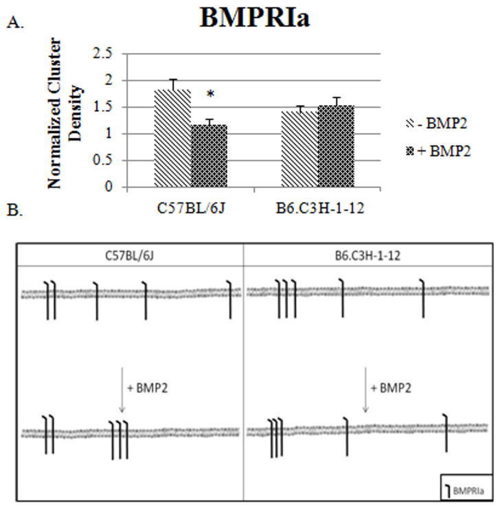
Figure 3. BMP2 stimulation led to a decrease in the clustering of BMPRIa on the cell surface of BMSCs isolated from C57BL/6J mice and no change for B6.C3H-1-12 mice.
BMSCs were isolated from 8 week old female C57BL/6J and B6.C3H-1-12 mice and stimulated or not stimulated with BMP2 (40nM). Cells were fixed using acetone/methanol and labelled for BMPRIa using a polyclonal goat BMPRIa antibody (Santa Cruz) followed by a secondary donkey anti goat Alexa 546 (molecular probes). High magnification images of flat regions of the plasma membrane were collected using a confocal microscope and the CD of BMPRIa was calculated. A) In the C57BL/6J the CD decreased with BMP2 stimulation. There was no change in CD in B6.C3H-1-12 mice with BMP2 stimulation. B) The summary of the CD results for BMSCs isolated from the C57BL/6J and B6.C3H-1-12.* Indicates significance as detected by one tailed student t test (p<0.05).

