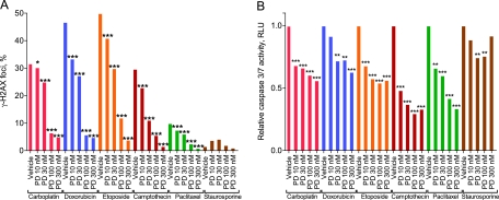
Figure 1.
Ability of pharmacological quiescence to protect against chemotherapy-induced γ-H2AX formation and apoptosis. The tHDF cells were treated with varying concentrations of PD0332991 (PD) before treatment with 100 μM carboplatin, 1 μM doxorubicin, 5 μM etoposide, 156 nM camptothecin, 250 nM paclitaxel, or 1.5 nM staurosporine. The tHDF cells were then assayed for A) γ-H2AX formation and B) caspase 3/7 activation. γ-H2AX formation data measured by flow cytometry represent at least 20 000 gated events. Caspase 3/7 data are presented as the average ratio from a single experiment of 1000 cells per well in quadruplicate. P values were calculated by a two-sided χ2 test (γ-H2AX formation data) or one-way analysis of variance (caspase 3/7 data) with Bonferroni correction for multiple comparisons. All data are representative of three independent experiments. *P < .05, **P < .01, ***P < .001. RLU = relative light units; tHDF = telomerized human diploid fibroblast.

