
Figure 2.
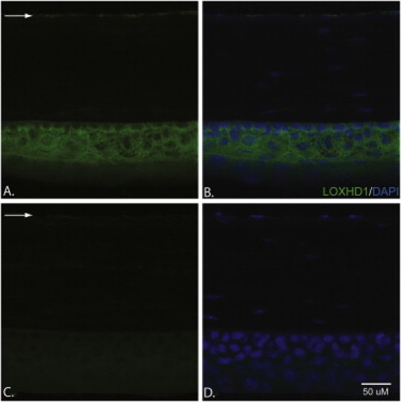
Immunohistochemical Analyses of LOXHD1 in 20 μm Cryosections of Mouse Cornea
(A and B) A slide stained with the LOXHD1 antibody (A) and a slide stained with both DAPI and the LOXHD1 antibody (B). LOXHD1 is present both in the corneal epithelium and in the endothelium; however, the level of LOXHD1 is much higher in the epithelium than in the endothelium.
(C and D) A slide stained with a LOXHD1 antibody +50× peptide (C) and a slide stained with both DAPI and a LOXHD1 antibody +50× peptide (D). The expression in the corneal epithelium and endothelium could be competed away with a commercially available LOXHD1 peptide.
The arrows point to the LOXHD1 present in the corneal endothelium.