
Figure 3.
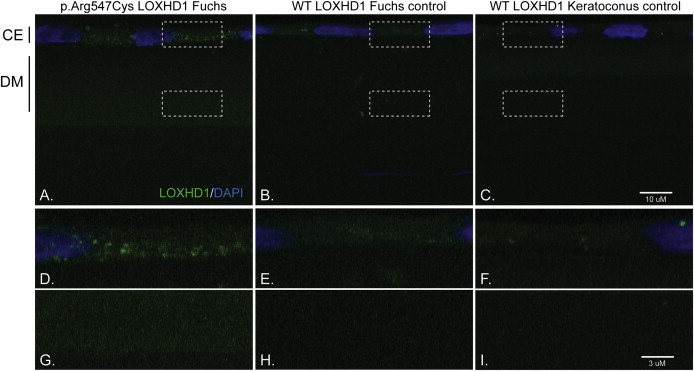
Immunohistochemical Analyses of LOXHD1 in Human Corneal Endothelium and Descemet Membrane
(A) Fuchs-affected individual harboring the c.1639C>T (p.Arg547Cys) LOXHD1 mutation.
(B) Fuchs-affected individual negative for any causal LOXHD1 mutation.
(C) Keratoconus-affected individual negative for Fuchs corneal dystrophy (note: panels D, E, F, G, H, and I are enlarged images of the boxed areas in panels A, B, and C). Aggregates of LOXHD1 are noted in the corneal endothelium and throughout the thickened Descemet membrane of a Fuchs patient harboring the c.1639C>T (p.Arg547Cys) LOXHD1 mutation. Few protein deposits in the corneal endothelium (“CE”) and Descemet membrane (“DM”) are seen in a Fuchs patient negative for any causal LOXHD1 mutation and in the patient transplanted for keratoconus. Note: The gain (exposure) is an order of magnitude higher in (C) than in (A) and (B).