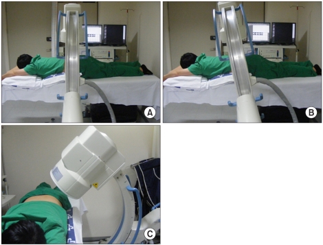
Fig. 2.
The positioning of the patient and C-arm are similar to lumbar discography. (A) The patient is placed in the prone position on a fluoroscophy table top padded to provide flattening of the lumbar lordosis. (B) The targeted disc's endplates are aligned as for discography with appropriate caudal or cranial tilt of the C-arm. (C) The beam is then rotated so that the lateral surface of the superior articular process (SAP) bisects the interspace, typically 40-45 degrees off the AP axis.

