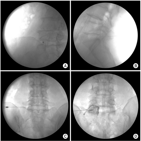
Fig. 3.
Retrodiscal injection L5-S1. (A) In oblique view, needle tip is advanced slowly and cautiously past the SAP lateral surface. (B) The lateral radiography should also be used while advancing past the SAP to minimize the risk of the penetration, while the resistance to the needle advancement is also used as sign to stop. (C) The AP view will most often demonstrate the tip in the interpedicular line. (D) A small amount of contrast is used to confirm epidural spread.

