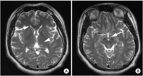
Fig. 1.
Brain magnetic resonance imaging (MRI) scans of a 24-year-old man with ataxia, dysarthria, and ophthalmic symptoms. T2-weighted MR images show no definite structural abnormalities or signal changes in the periventricular white matter (arrow), the medial thalamus (arrow head) (A), or the mamillary bodies (arrow) (B).

