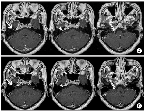
Fig. 2.
Contrast internal auditory canal MRI. (A) Contrast-enhanced axial T1-weighted image (onset) shows enhancement in the geniculate ganglion (arrow), cisternal and in ternal auditory canal segment of facial and vestibulocochlear nerve (arrowhead), and cisternal segment of the glossopharyngeal and vagus nerve (open arrow). (B) Contrast-enhanced axial T1-weighted image (2 months after onset) shows decreased enhancement in the geniculate ganglion (arrow), cisternal and internal auditory canal segments of facial and vestibulocochlear nerves (arrowhead), and cisternal segment of the glossopharyngeal and vagus nerves (open arrow).

