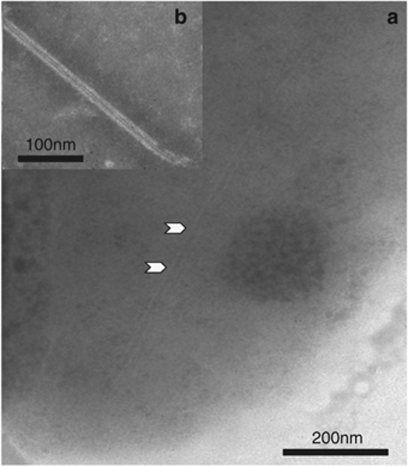
Figure 3.
Transmission electron photomicrograph of Vc450 T6SS tubular structure. Vc450 cell stained with 0.5% sodium phosphotungstic acid, pH 6.8. (a) A VipA/VipB-like T6SS tubular structure, similar to that described for V. cholerae, is evident in the cytoplasm (black arrowheads). (b) A Vc450 VipA/VipB-like T6SS tubular structure found outside of a cell.

