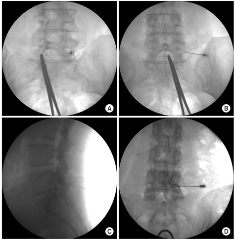
Fig. 5.
Subpedicular approach of the L5 nerve root. (A) In oblique view, needle tip lies directly inferior to the pedicle and inferolateral to the pars interarticularis. (B) The anterior-posterior view showing the proper location of the needle at the base of pedicle. (C) The lateral radiography should also be used while the needle is advanced until the needle tip is at the anterior and superior aspect of intervertebral neural foramen. (D) A small amount of contrast is used to confirm epidural spread.

