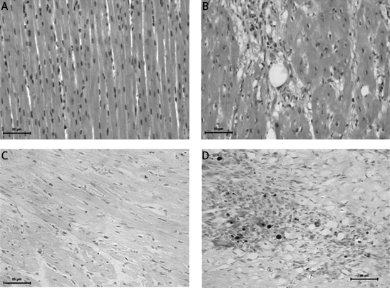
Figure 4.
HE staining of myocardium and apoptosis before and after CME (400×). A – Posterior myocardium after CME. B – Anterior myocardium after CME. Microsphere is at the middle. The microsphere is surrounded by inflammation and infarction. C – Posterior myocardium after CME. No apoptosis is observed. D – Anterior myocardium after CME. Dark nuclear indicates apoptosis after CME

