Figure 3.
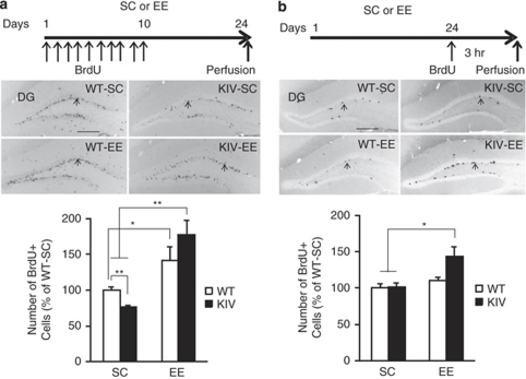
Survival and proliferation of progenitors in brain-derived neurotrophic factor knock-in IV (BDNF-KIV) mice and effects of enriched environment (EE). Top panels: BrdU labeling paradigm. Middle panels: representative images displaying BrdU positive (+) cells (arrows) from the four groups. Lower panels: quantification of BrdU+ cells in the dentate gyrus (DG). (a) Number of surviving progenitor cells (3 weeks). Note the decreased number of surviving BrdU+ cells in KIV-standard condition (SC) compared with wild-type (WT)-SC and the significant increase in the number of BrdU+ cells in KIV-EE mice compared with both KIV-SC and WT-SC (18–20 DG in 9–10 sections from each brain, n=5 per group). (b) Number of proliferating cells (3 h). Note the significant increase in the number of BrdU+ cells in KIV-EE compared with KIV-SC and WT-SC (8–10 DG in 4–5 sections from each brain, n=3 per group). Results are expressed as % of WT-SC±s.e. Scale bar in (a) and (b)=250 μm. *P<0.05, **P<0.01.

