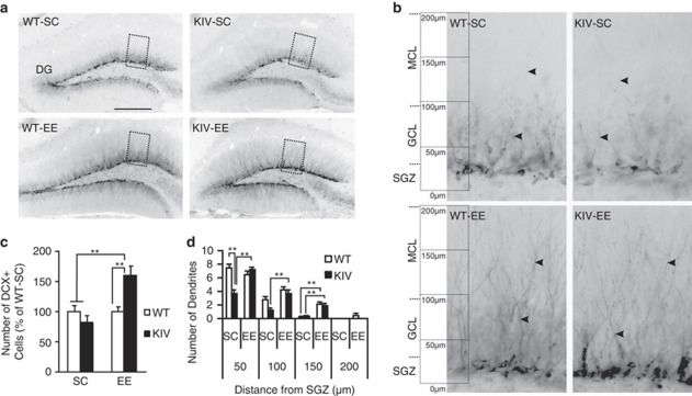
Figure 4.
Doublecortin-positive (DCX+) cells and dendritic extension in the dentate gyrus (DG) and effect of enriched environment (EE). (a) Representative images showing DCX+ cells and their dendrites from the four groups. Scale bar=250 μm. (b) Magnified view of boxed area in (a) showing DCX+ cells and their dendrites (arrowheads). (c) Quantification of DCX+ cells in the DG. Note the significantly increased number of DCX+ cells in knock-in IV (KIV)-EE mice compared with KIV-standard condition (SC) and wild-type (WT)-SC groups. Results are expressed as % of WT-SC±s.e. (d) Quantification of the extension of apical dendrites of DCX+ cells into the molecular cell layer of the DG. Note the significantly reduced numbers of dendrites in KIV-SC compared with WT-SC at 50 μm from the subgranular zone (SGZ), which was rescued by EE. EE increased the number of dendrites of both genotypes at 150 μm from the SGZ. (Two DG from each brain, n=4 per group). Results are expressed as mean±s.e. **P<0.01.

