
Fig. 1.
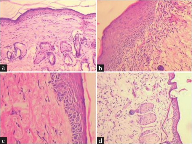
Photomicrophotographs of sections of mouse skin at 5 days post wounding
Representative microphotographs of section of mouse skin at 5 days post wounding which received twice daily showing (a) normal wound healing in vehicle treated group (b) vascular proliferation with presence of fibroblasts in VEGF (1 μg/g) treated group (c) healing with dense collagenation in dermis of TP (1 mg/g) treated group and (d) inflammation and edema with poorly laid collagen in TP (4 mg/g) treated group. [H and E ×100 (1a and 1d); H and E ×200 (1c and 1b)].