Figure.
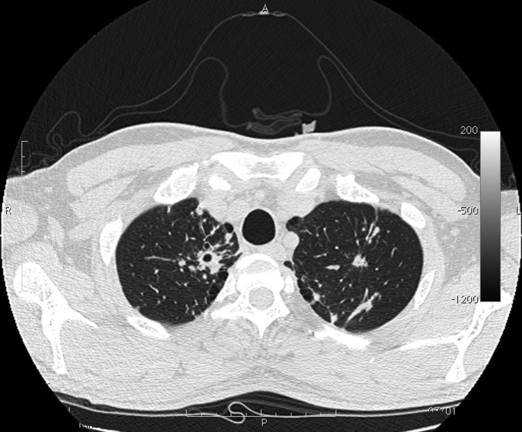
Computed tomography image of chest of patient with tuberculosis after anti–hepatitis C virus therapy. A parenchymal distortion 32 mm in diameter is shown in the upper right lung with initial central excavation 10 mm in diameter. Similar lesions 8 mm in diameter without central excavation are shown in the upper left lung.

