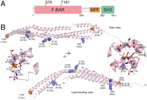
Fig. 2.
Location of S76 and T181 on the syndapin I F-BAR. (A) Schematic location of S76 and T181 phosphosites in the domain structure of rat syndapin I. (B) Cartoon of the human syndapin I F-BAR (PDB ID code 3HAI). The side view (Top) and lipid-binding view (Bottom) of monomer are shown. The homologous human residues for S76 and T181 phosphosites are shown in orange spheres, and the basic residues involved in lipid binding are shown in blue spheres. Numbering for the rat sequence residues are in brackets. The close-up of S76 and T181 show potential hydrogen bonds (dashed line) with D276 and D280, and E183 and Q184 residues, respectively. Note that the sequences of rat and human syndapin I differ such that rat S76 is S79 in humans, and rat T181 is T184 in humans.

