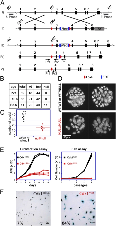
Fig. 1.
Generation and analysis of Cdk1 conditional knockout mice and cells. (A) The Cdk1 genomic locus (I) was modified in ES cells with the targeting vector (II) shown. An FRT-flanked neomycin-selection cassette was introduced along with LoxP recombination sites (red triangles) on both sides of exon 3, which generated a mutant Cdk1 locus (III). For Southern blot analysis, 5′ and 3′ probes located outside of the targeting vector were used, after an EcoRV (RV) digest, resulting in a 31,206-bp (recombinant) and a 9,110-bp (5′) or 20,300-bp (3′) fragment for wild type (Fig. S1A). Upon expression of FLP recombinase, the neomycin cassette was removed, and only the LoxP sites remained in the locus (IV, Cdk1FLOX). The levels of Cdk1 protein expression were similar in Cdk1FLOX and Cdk1WT mice (Fig. S1B). After Cre recombinase expression, exon 3 was excised (V), which resulted in deletion of Cdk1 and a frame shift. PCR genotyping primers are indicated (Pr1, -2, and -3), and the sequences can be found in Table S1. (B) To generate Cdk1 knockouts, Cdk1FLOX mice were crossed with β-actin–Cre mice expressing Cre recombinase ubiquitously in all tissues, including germ line. The resulting Cdk1WT/NULL mice were interbred, and the offspring was analyzed at weaning (P21), midgestation (E10.5), or blastocyst (E3.5) stage. (C and D) Cdk1NULL/NULL blastocysts were visualized by Hoechst staining of their nuclei followed by fluorescence microscopy. Comparison of Cdk1-deficient blastocysts with heterozygous or wild-type littermates indicated a reduced number of cells (C); however, their nuclei were larger in size (D). (E and F) Three independent MEF lines (Fig. S1 C–H) were treated with 4-OHT to induce Cdk1 knockout, and their proliferative potential was monitored by 3T3 and alamarBlue proliferation assays for several passages or days, respectively (E). Deletion of Cdk1 resulted in a rapid arrest of cellular proliferation and premature onset of cellular senescence that was detected by senescence-associated β-gal staining (F). Therefore, cells lacking Cdk1 cannot proliferate but instead enter a senescent state and survive in culture medium.

