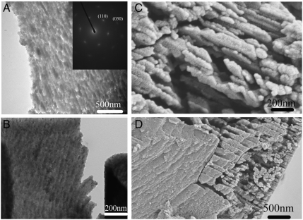
Fig. 2.
(A) TEM image of a thin section of a sea urchin spine (floated onto water). Inset: SAED pattern indicating the section was cut perpendicular to the [001] direction of calcite. (B) TEM image of a thin section of a sea urchin spine (floated onto ethylene glycol). (C), (D) Images of fractured sea urchin spine etched in water overnight.

