Fig. 1.
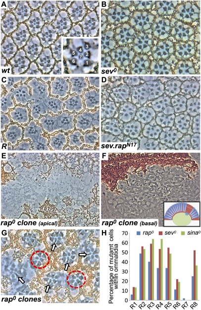
rap is required for R7 specification. (A) A tangential section through a wild-type eye. Inset shows large rhabdomeres of R1–R6 surrounding the central small R7. (B) A tangential section through a sev0 eye shows only the six outer rhabdomere cells. (C) A tangential section through a Roughened eye. Many ommatidia lack R7 cells. (D) sev.rapN17 phenocopies Roughened; many R7 cells are absent. (E) A section through a large rap0 clone marked by the absence of pigment. (F) Apically, clones lack photoreceptors, which are found lying below the retina and above the lamina nuclei level. Inset shows a schematic representation with the upper white dotted box indicating the region shown in E and the lower box indicating the area shown in F. (G) A tangential section through an eye containing many late-induced rap0 clones. Normally constructed ommatidia containing a mixture of mutant and wild-type cells are found. R7 cells are almost invariably pigmented (rap+) (arrows). Additionally, many ommatidia lack R7 cells (red circles). (H) Histogram showing the percentage of mutant photoreceptors found in rap0 (blue bars), sev0 (red bars), and sina0 (green bars; adapted from ref. 27) mosaic ommatidia.

