Fig. 2.
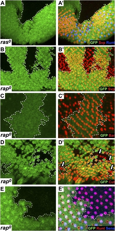
A comparison of ras0 and rap0 mutant clones in third-instar eye discs. In all images, the clones are indicated by the absence of GFP (green) and are circumscribed by a dashed line. (A and A′) A ras0 clone shows no differentiating ommatidia. (B and B′) rap0 clones show extensive Svp staining (red), which labels the R1/3/4/6 photoreceptors. (C and C′) rap0 clones contain many cells expressing Bar (red), which selectively marks R1/6 cells. (D and D′) rap0 clones show the cone cell marker Cut (red; arrows). (E and E′) rap0 clones stained for Runt (red) and Sens (blue). Cells expressing Sens and Runt (R8 cells) populate the clone, but Runt-positive/Sens-negative R7 cells are only found in the wild-type tissue (asterisks).

