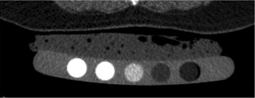
Figure 2d:
Volumetric quantitative CT scans of the lumbar spine (L3 vertebral body) obtained in a 64-year-old man show (a) axial source image and (b) sagittal and (c) coronal reformation. In addition a five-element calibration phantom is shown in d, which was used to transform Hounsfield units into milligrams hydroxyapatite per milliliter (BMD).

