
Figure 6:
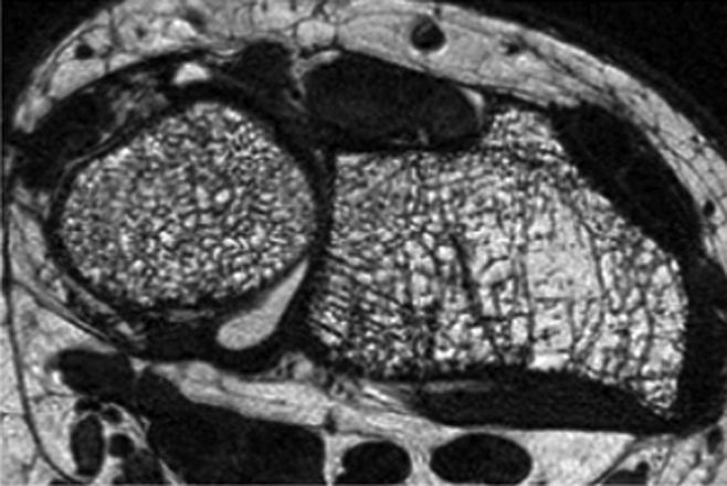
Distal radius high-spatial-resolution MR image (voxel size: 0.156 × 0.156 × 0.5 mm3) obtained at 3.0 T with a fully balanced steady-state free precession sequence and a transmit-receive quadrature wrist coil in a 55-year-old postmenopausal woman. Heterogeneity of trabecular bone architecture and focal loss of trabecular bone are well demonstrated.