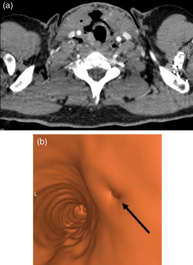
Figure 1:

(a) CT image demonstrate extensive subcutaneous emphysema and the defect in cervical trachea. (b) Virtual bronchoscopy demonstrates an elliptic defect in right antero-lateral tracheal wall (black arrow) at the level of the fourth tracheal ring (L = left).
