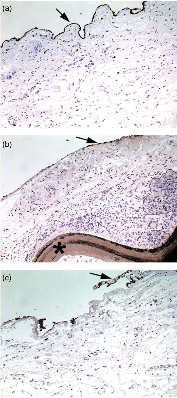
Figure 3:

Representative picture of the epicardial coverage by mesothelial cells (arrow). (a) Cova™ CARD sheep; (b) Preclude® (star) sheep; (c) control sheep. Immunostaining of the mesothelial cells by an antibody against cytokeratins AE1/AE3. Original ×10. Note the continuous cell lining in the Cova™ CARD sheep whereas it is interrupted or absent in the Preclude® and control sheep, respectively.
