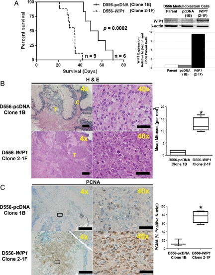
Fig. 4.
High WIP1 expression promotes growth of intracerebellar xenografts of human medulloblastoma cells in vivo; 5 × 10⁵ D556-pcDNA (Clone 1B) or D556-WIP1 (Cone 2-1F) cells were injected into the cerebellum of 3–6-month-old SCID/Beige mice. (A) Kaplan-Meier plot comparing survival among mice bearing D556-WIP1 and D556-pcDNA xenografts (P = .0002). Right panel, WIP1 expression by Western blotting in parental D556, D556-pcDNA (Clone 1B), and D556-WIP1 (Cone 2-1F) cells with β-actin as a loading control. Below Western blot, quantification of WIP1 expression relative to β-actin and expression in parental D556 cells. (B) Representative hematoxalin and eosin (H&E)-stained saggital sections of tumors from symptomatic SCID/Beige mice xenografted with D556-pcDNA (Clone 1B) or D556-WIP1 (Clone 2-1F) cells. Right panel, quantification of mitotic figures in H&E-stained sections of D556-pcDNA (Clone 1B, n = 4) or D556-WIP1 (Clone 2-1F, n = 4) xenografted tumors (*P = .0003). (C) Representative immunohistochemistry for the marker of proliferation, PCNA (n = 4 for each group). Right panel, Quantification of nuclear staining for PCNA in D556-pcDNA (Clone 1B) or D556-WIP1 (Clone 2-1F) xenografted tumors (*P = .001). The middle line in each box represents the median value for each group. Whiskers represent the minimum and maximum values in each group. Bars in photomicrographs measure 500 μm (4×) or 100 μm (40×).

