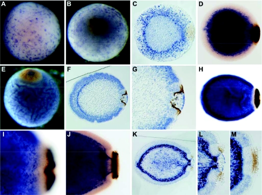
Fig. 3.
Developmental expression of Aq-Cry1. (A,B,E) Whole-mounts; (C,F,G,K,L,M) sections; (D,H,I,J) cleared whole-mounts; (B,E) posterior pole facing up; (C,D,F–M) posterior pole to right. (A,B) In early gastrula and spot stages, Aq-Cry1 expression is evident in maternal follicle cells on the surface of the embryo. (C,D) After gastrulation, at the spot stage, embryonic Aq-Cry1 expression is strongest in a population of cells scattered throughout the outer region of the embryo. (E,F) During ring formation, the gene is upregulated in presumptive epithelial-like cells and in a population of cells inside the pigment ring. (G) Higher magnification of the embryo shown in F, showing expression diminished in cells adjacent to the outer part of the ring. (H) Excluding cells adjacent to the pigment ring, expression in the outer layer continues in early larvae (pre-release). (I) Higher magnification of the embryo shown in H. (J,K) In hatched, swimming larvae, Aq-Cry1 is expressed in sub-epithelial cells and cells located inside the ring, and in the epithelial-like cells a in a pattern similar to that observed at earlier stages. (L,M) Higher magnifications of the embryo shown in K, sectioned through the middle of the ring (L) or the edge of the ring (M).

