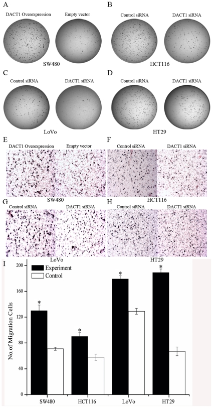
Figure 3. DACT1 enhances anchorage independence and the migratory potential of colon cancer cells.
(A) Soft agar assay in DACT1-transfected SW480 cells after 14 days of incubation. (B, C, D) Soft agar assay in siRNA-transfected HCT116, LoVo and HT29 cells after 14 days of incubation. (E) Representative images are shown for the overexpression of DACT1 in stably transfected SW480 cells. Cells were seeded in a transwell chamber and allowed to migrate across the chamber toward cell-specific conditioned medium for 24 h. Photomicrographs of stained migrating cells were taken under brightfield illumination (20×). (F, G, H) Representative images are shown for DACT1 siRNA-transfected HCT116, LoVo and HT29 cells. Cells were seeded in a transwell chamber and allowed to migrate across the chamber toward cell-specific conditioned medium for 24 h. Photomicrographs of stained migrating cells were taken under brightfield illumination (20×). (I) Quantification of migration assay. Results were obtained from three separate experiments each performed in triplicate. Migration was determined by counting cells in six random microscopic fields per well (mean ± SEM; *p<0.05 versus control group cells).

