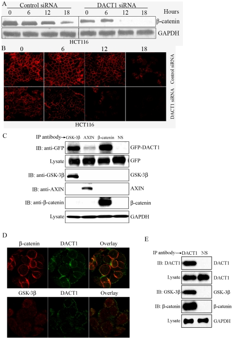
Figure 8. DACT1 interacts with β-catenin and components of the β-Catenin destruction complex to stabilize β-catenin.
(A) Western blot assays in HCT116 cells were performed to determine β-catenin stability. (B) Immunofluorescence assays in HCT116 cells were performed to determine β-catenin stability. (C) Cell lysates from SW480 cells transfected with GFP-DACT1 were subjected to immunoprecipitation (IP) with anti-axin, anti-GSK-3β, or anti-β-catenin antibodies. Immunocomplexes were resolved by SDS-PAGE and subjected to Western blot analyses with an anti-GFP antibody. Blotting with an anti-GAPDH antibody showed equal loading. (D) Subcellular co-localization of endogenous DACT1 and β-catenin, DACT1 and GSk-3β in HT29 cells. (E) Cell lysates from HT29 cells were subjected to immunoprecipitation (IP) with an anti-DACT1 antibody. Immunocomplexes were resolved by SDS-PAGE and subjected to Western blot analyses with anti-GSK-3β, or anti-β-catenin antibodies. Blotting with an anti-GAPDH antibody showed equal loading.

