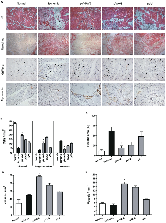
Figure 6. Morphometric analysis of limb muscles.
The gastrocnemius muscle was collected from mice after four weeks of gene therapy. Tissue samples were stained with HE (A) and used to quantify necrotic, regenerative and normal areas (B). The sham-operated and non-ischemic groups showed no difference in their results and are denoted here as normal. Fibrotic area, capillary density and mature vessel density were determined after staining with Picrosirius (C), Griffonia (D) and alpha-actin antibody (E), respectively. Bar = 50 µm. * p<0.05. ▴, Infiltrated mononuclear cells; X, adipocytes; →, capillary; In the figure B, pVHAVI was different statistically in comparison to all groups.

