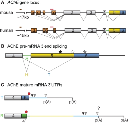
Figure 1.
Schematic diagrams depicting the mouse and human AChE genes, alternative splicing options and cis-element locations. (A) Mouse and human AChE genes, including the leader exons identified in brain tissues. Noteworthy is the 5′ regulatory region that harbors alternate exons 1a–e in mouse and 1a–d in human (orange boxes), distal enhancer glucocorticoid response element (brown line) and proximal muscle-specific enhancer (pink line). Gray boxes and black lines represent constitutive exons and introns, respectively. (B) Alternative splicing at the 3′ end of AChE pre-mRNA produces tissue-specific R (green), H (yellow) and T (blue) variants. Approximate locations of the U-rich regulatory sequence, SC35 and ASF/SF2 splicing factor binding sites are denoted by black, white, and gray stars, respectively. (C) The 3′ untranslated regions (3′ UTRs) of mature T and R AChE variants and the alternative polyadenylation-dependent extended regions. The AChEH variant is not shown since it is not significantly expressed in neurons. Symbols represent the PBE (black triangle), ARE (gray triangle), miR-132 binding (white triangle), translation stop (red octagon), and polyadenylation signal (p(A)) sites. 4′ signifies a pseudo-intron that contains the AChER translation termination site. Whether E6 and the downstream region are included in AChER transcripts is unknown (?).

