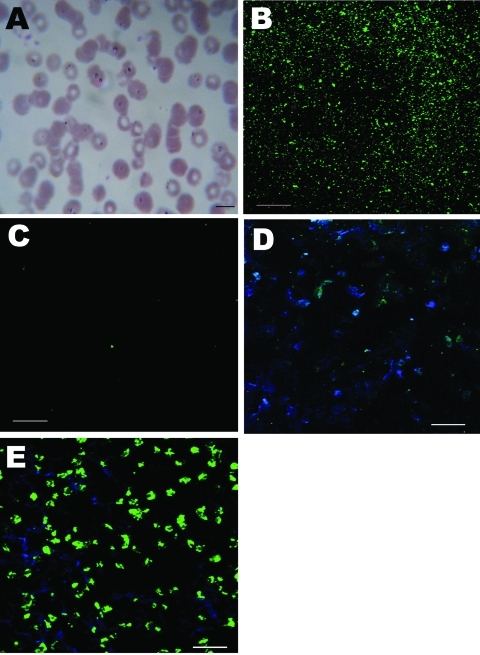
Figure 1.
Morphologic appearance of infected erythrocytes of a 57-year-old woman in China and immunofluorescent antibody test results. A) Giemsa-stained thin blood smear showing erythrocyte infected with multiple ring forms (arrowhead). Scale bar = 10 µm; original magnification ×100. B) Patient serum reactive against Colpodella antigen. Scale bar = 20 µm; original magnification ×40. C) Healthy control serum not reactive against Colpodella antigen. Scale bar = 20 µm; original magnification ×40. D) Patient serum not reactive (green fluorescence) against Babesia microti antigen. Scale bar = 20 µm; original magnification ×40. E) B. microti–infected mouse serum reactive against B. microti antigen. Scale bar = 20 µm; original magnification ×40.

