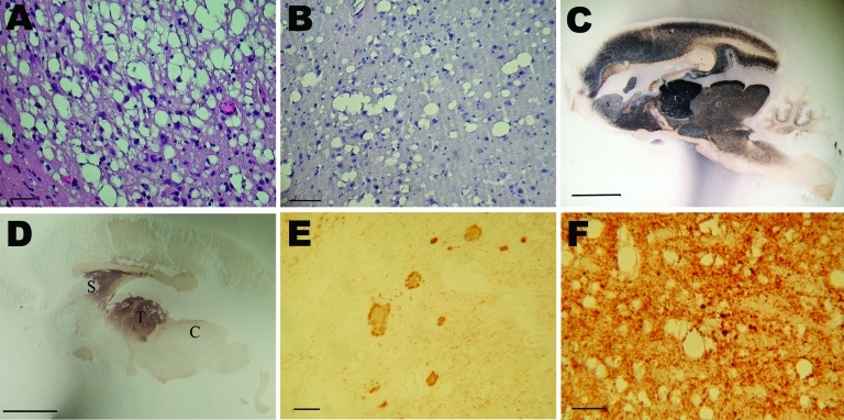
Figure 2.
Histopathologic and disease-associated prion protein (PrPd) immunodetection in the brain of 2 mouse lemurs after intracerebral (5 mg) or oral (50 mg) inoculation with a cattle-derived L-type bovine spongiform encephalopathy isolate. A, B) Spongiosis in the striatum; scale bars = 30 μm. C, D) Paraffin-embedded tissue blot analysis of sagittal brain section; scale bars = 500 μm. E, F) PrPd immunodetection; scale bars = 30 μm. Analyses in C–F were performed by using the 3F4 monoclonal antibody against PrP. C, colliculus; S, striatum; T, thalamus.

