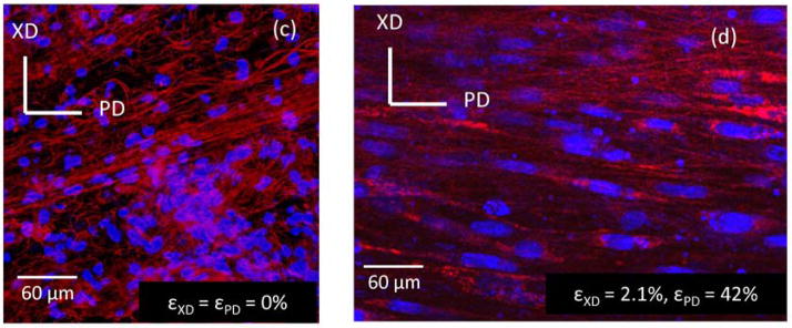
Fig. 3.
Fluorescent microscope images showing cell-scaffold constructs made via electrospinning PEUU and concurrently electrospraying rat vascular smooth muscle cells onto a rotating mandrel. (Left, c) Constructs in a static (non-strained) environment show rounded cell nuclei (blue) and random fiber orientation (red). (Right, d) Constructs under biaxial strain show elongated cell nuclei and PEUU fiber alignment, demonstrating enhanced mechanical properties of electrospun polymers compared with natural polymer systems. Reproduced with permission from [114]

