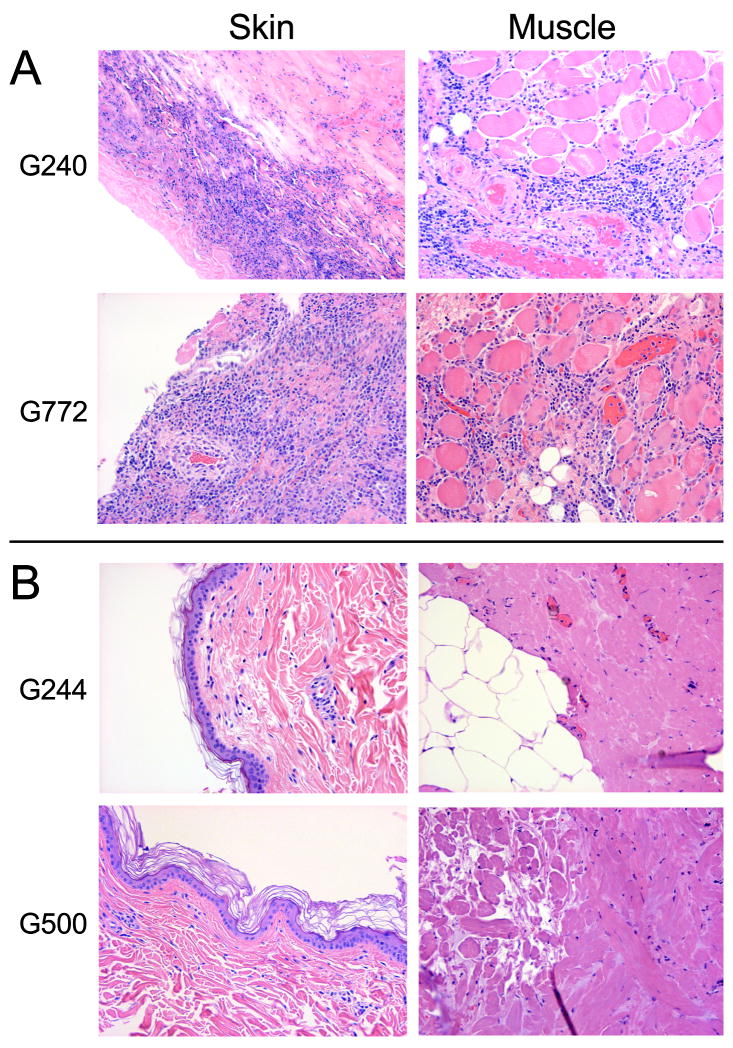
Figure 2. Histology of transplanted skin and muscle tissue from marrow donors and mixed chimeric recipients.
Recipient grafts in both marrow donors, G240 and G244 show dense lymphocytic infiltrates in skin and muscle on day 21 and 31 respectively; consistent with rejection (A). Marrow donor grafts in both mixed chimeras G244 and G500 showed normal skin and muscle histologies on days 167 and 118, respectively (B). Slides were photographed at 200X magnification.

